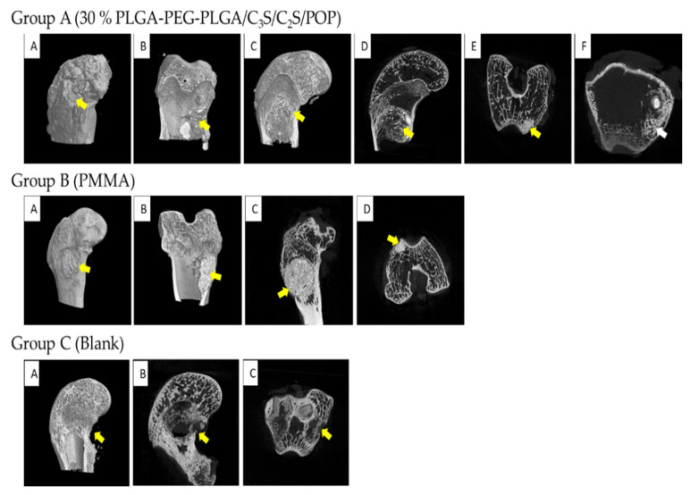
Figure 14.
MICRO-CT results of femoral condyles and implant material 6 weeks after operation. Group A (30% PLGA-PEG-PLGA/C3S/C2S/POP): (A–C) three-dimensional reconstruction; (D–F) sagittal, coronal, and horizontal, respectively. The yellow arrow pointed to the composite bone cement filler material and the white arrow pointed to the new bone trabecula. Group B (PMMA): (A,B) reconstructed in three dimensions; (C,D) sagittal and coronal, respectively. The yellow arrow refers to PMMA bone cement filling material, which had a clear boundary with the surrounding bone, and no obvious new bone trabecula was found. Group C (Blank): (A) three-dimensional reconstruction map, (B) sagittal plane, and (C) coronal plane. Yellow arrow pointed to the defect of the femoral condyle, and no healing was observed.

