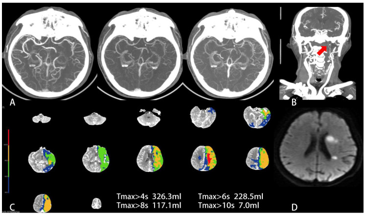
Figure 2.
Baseline mCTA, CTP and follow-up DWI in a 70-year-old man with wake-up stroke. The time from last known well to CT scan was 12.5 h. The baseline NIHSS score was 7 and the ASPECTS score on NCCT was 7. (A) mCTA axial MIP shows robust pial collateral vessels within the left temporal parietal lobe and only a one-phase delay in the filling of peripheral vessels. The mCTA collateral score was 4 (favorable collateral status). (B) Coronal reconstruction of neck CTA MIP shows severe stenosis/occlusion of C1–4 segment of left internal carotid artery (red arrow). (C) Tmax map shows the severe hypoperfusion area (red) defined Tmax > 10 s was 7 mL and a hypoperfusion area (light green) defined Tmax > 6 s was 228.5 mL, while HIR (=0.03) was very low. (D) This patient underwent a successfully EVT and had a FIV of 8.6 mL on the left basal ganglia and paraventricular region on DWI. The 90 d mRs score of this patient was 2 (favorable functional outcome).

