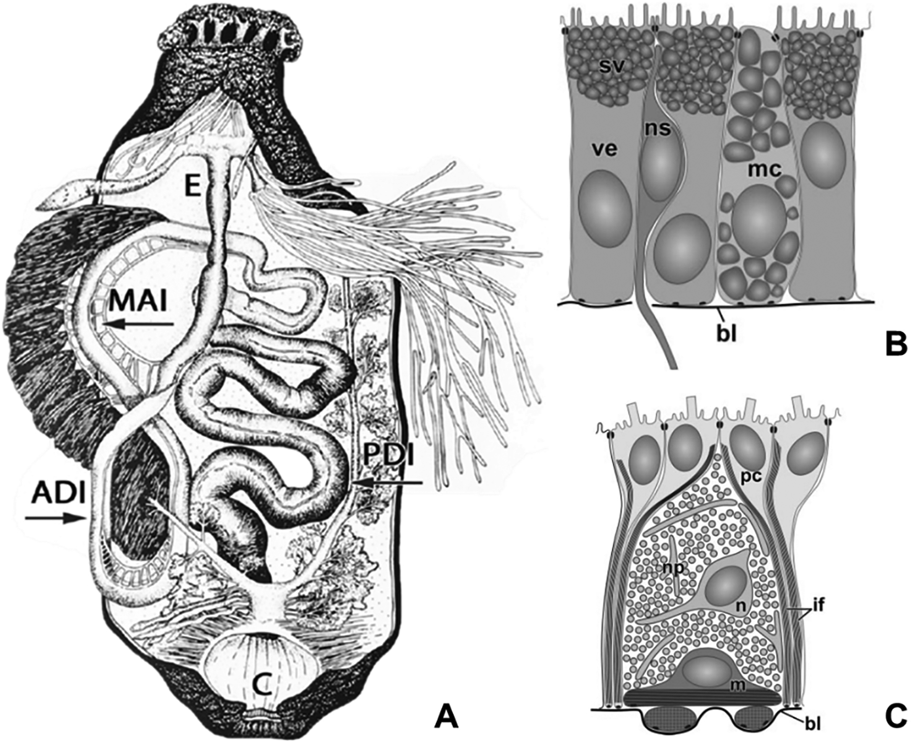
Fig 2. Anatomical organization and tissue architecture of the sea cucumber digestive.

(A) Visceral anatomy of sea cucumber. The intestine is subdivided into the anterior descending small intestine, ascending small intestine, and the posterior descending large intestine. Abbreviations: short esophagus (E); anterior descending intestine (ADI); medial ascending intestine (MAI); posterior descending intestine (PDI). (B) Tissue organization of the luminal epithelium and (C) mesothelium found throughout the intestine of H. glaberrima. Abbreviations: basal lamina (bl); bundles of intermediate filaments in peritoneocytes (if); myoepithelial cell (m); mucocyte (mc); neuron (n); neurosecretory cell (ns); nervous plexus (np); peritoneal cell (pc); secretory vacuoles (sv); vesicular enterocyte (ve). Fig. 2 A adapted from García-Arrarás et al., 2019, Seminars in Cell & Developmental Biology 92, p. 47 and fig 2 B, C from Mashanov & García-Arrarás, 2011, Biological Bulletin, 221, p. 95. Copyright 2019 by Elsevier (2 A) and 2011 by The University of Chicago Press (2 B, C). Adapted with permission.
