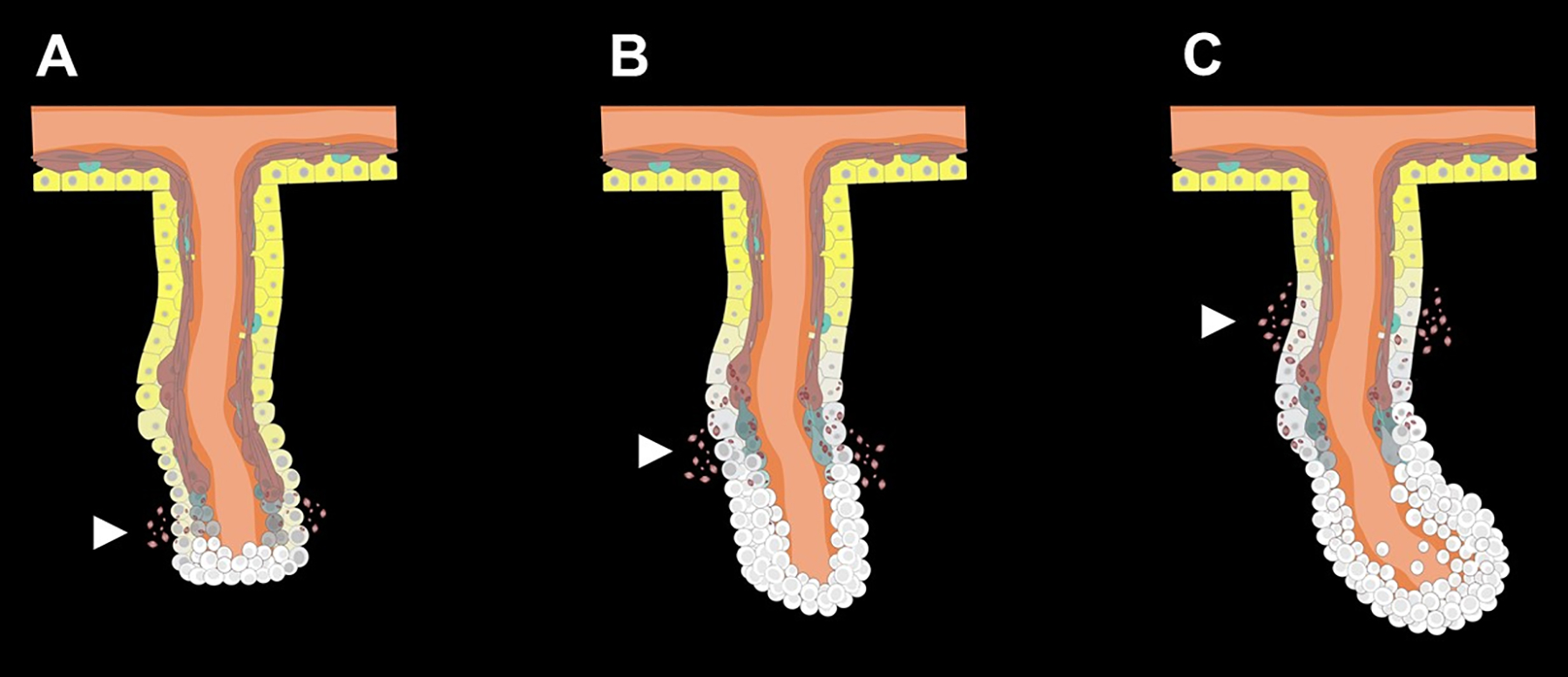
Fig 3. Cellular dedifferentiation and proliferation at the early stage of the regenerative process.

(A) Cells in the free end of the mesentery begin to dedifferentiate soon after evisceration. (B) Cells accumulate at the free edge of mesentery forming the early blastema-like structure. (C) As regeneration proceeds, dedifferentiation spreads along the mesentery and involves regions closer to the body wall, while cell proliferation remains mainly restricted to the blastema-like structure composed of the dedifferentiated cells. White arrows point out SLSs from the dedifferentiation process. White cells represent dedifferentiated cells originated from peritoneocytes (yellow) and muscle cells (reddish-brown) of the mesothelium. Connective tissue (orange) is positioned between the layers of the mesothelium and extends to the body wall (upper part).
