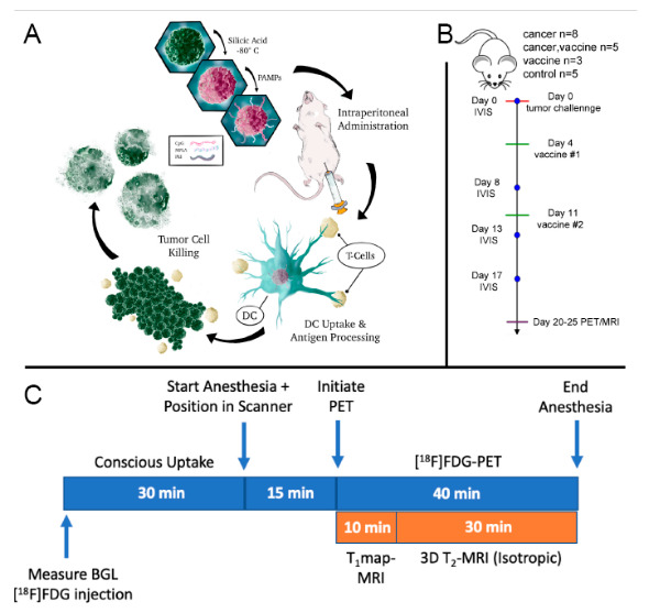
Figure 2.

Study design and underlying mechanism of action for cancer immunotherapy. (A) Schematic of vaccine preparation and in vivo-stimulated immune response. Briefly, BR5-Akt cells were cryo-silicified and then coated with PEI followed by CpG and MPL for intraperitoneal administration on days 4- and 11-post tumor challenge. The schematic shows uptake of the vaccine by dendritic cells (DC), antigen presentation to T cells, and tumor cell killing. (B) Timeline for tumor challenge, vaccination, bioluminescence imaging (IVIS), and PET/MRI. (C) PET/MRI scan protocol. Animals were fasted for 3 h and blood glucose levels (BGL) were measured prior to conscious [18F]FDG administration and simultaneous PET/MRI.
