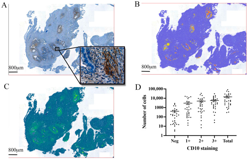
Figure 1.
CD10 and the automated identification of endometrial stromal cells in excised endometriotic tissue. (A) Mouse anti-CD10 antibody-labelled endometrial stromal cells in the excised tissue, revealing multiple endometriosis foci. (B) Automated cell detection was performed with QuPath software over the entire excised lesion. (C) Pixel smoothing identified the size of each endometriosis foci, each individual cell within the lesion, and the relative staining intensity of each cell. (D) Total cell counts showed the number of stromal cells identified by automated software and the number of cells that were considered either negative for CD10 staining, lightly (1+), moderately (2+), or heavily (3+) immunoreactive for CD10.

