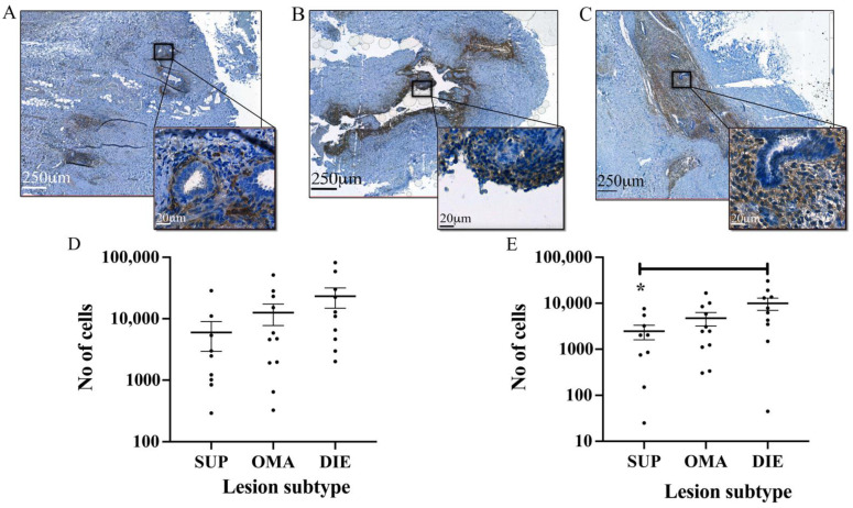
Figure 2.
CD10-positive stromal cells in endometriotic lesions of different subtypes. The CD10 antibody and automated analysis of images using QuPath identified endometriotic stromal cells in lesions from (A) SUP, (B) OMA, and (C) DIE. (D) The total number of stromal cells was lowest in SUP lesions followed by OMA lesions. DIE lesions contained the largest number of stromal cells. (E) There were significantly more stromal cells with heavy CD10 staining intensity (3+) in DIE lesions compared to SUP lesions. * p < 0.05.

