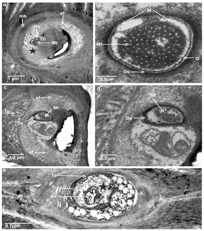
Figure 6.
TEM photograph explores the ultrastructures of the trichoid mechanoreceptor: (a) cross-section at the base of trichoid mechanosensillum, where tubular body (Tb) is visible; socket membrane or joint membrane (mb) (b) magnification microtubules (Mt) of the tubular body; (c) profound location of the mechanoreceptor, where tubular body is wider in this section of trichoid mechanosensillum; (d) enlargement and the shape of the tubular body; (e) tormogen cell (To) with the sensillum lymph cavity (marked star) and trichogen cell (Tr), dendrite sheath (Ds), line of granules (G), suspension fibre (sf), and membrane (M).

