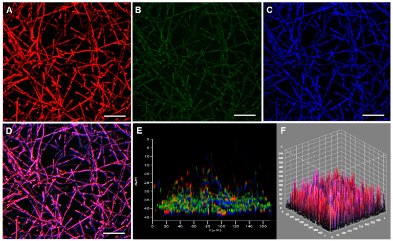
Figure 4.
Confocal microscopy of the biofilm formed by P. verrucosa cells on polystyrene. Conidia (1 × 10⁶) were incubated at 37 °C in polystyrene confocal plates containing RPMI medium. After 48 h, the fungal cells were stained with (A) FilmTracer SYPRO®, (B) TOTOTM, (C) calcofluor white M2R and (D) the staining combined. (E,F) 3-D reconstructions of the fungal biofilm. Bars, 20 μm.

