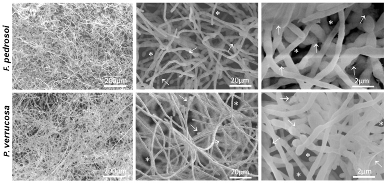
Figure 5.
Low-magnification SEM images of fungal biofilm on a polystyrene surface. Fungal cells (1 × 10⁶) were added to polystyrene cover slips and incubated for 72 h at 37 °C. Then, the cells were processed for SEM, as detailed in Material and Methods. Representative images of F. pedrosoi and P. verrucosa showed biofilm features as dense masses of mycelia containing inner water channels (asterisks) and extracellular matrix (white arrows).

