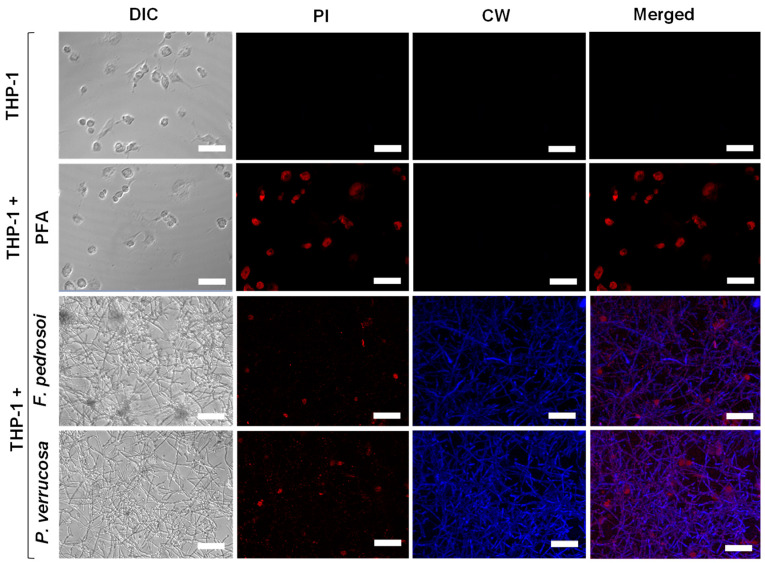
Figure 7.
Confocal microscopy of fungal biofilm formation on THP-1 cells. The human monocytic leukemia cell line (4 × 10⁵/mL) was added in 24-well cell culture plates containing RPMI medium supplemented with 80 nM PMA to differentiate macrophage cells, as detailed in Material and Methods. Representative images of THP-1 cells non-treated (control of viable cells), treated with paraformaldehyde (PFA, control of non-viable cells) or incubated for 48 h with (4 × 10⁶/mL) of F. pedrosoi and P. verrucosa. After co-culturing, the systems were washed with RPMI and incubated with propidium iodide (PI) and calcofluor white (CW). THP-1 cells damage by fungal biofilm formation was monitored using confocal differential interference contrast (DIC) and fluorescence microscopy. Bars, 50 μm.

