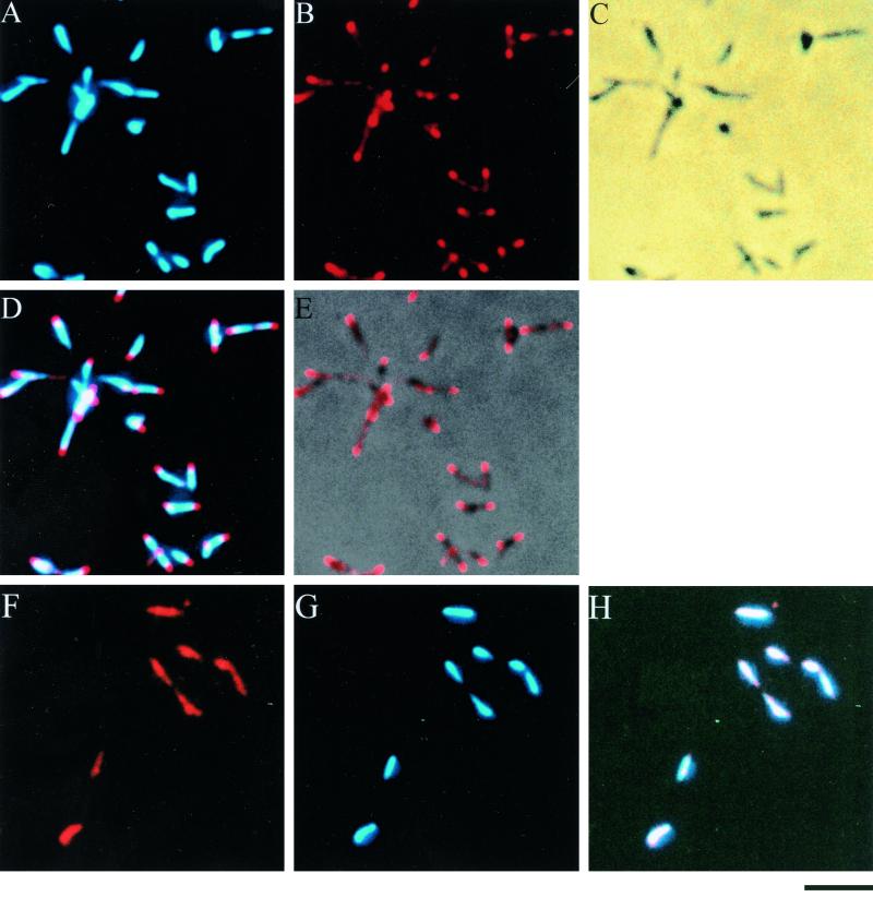
FIG. 1.
Subcellular localization of P1 adhesin (A to E) and HU protein (F to H) in M. pneumoniae. Cells were fixed, permeabilized, and stained with antibodies and DAPI. (A) DAPI-stained image; (B) anti-P1 antibody-stained image; (C) phase-contrast image; (D) merge of P1 and DAPI staining; (E) merge of P1 staining and phase-contrast image; (F) anti-HU antibody-staining image; (G) DAPI-staining image; (H) merge of HU and DAPI staining. Bar, 2 μm.

