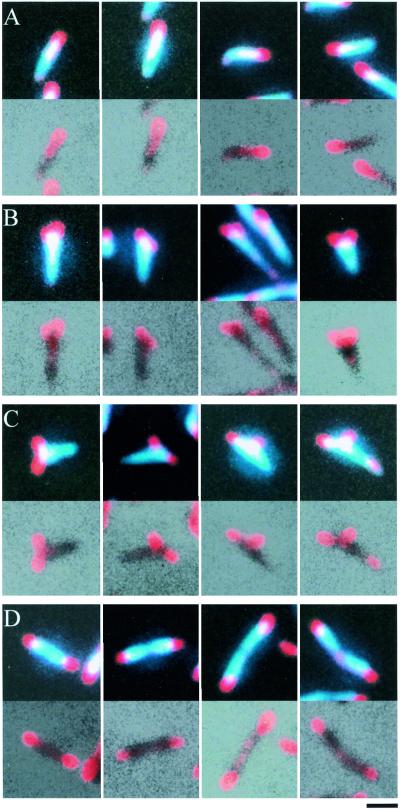
FIG. 2.
Cell image typing based on P1 localization. The upper and lower sections of each block show merges of P1 and DAPI staining and of P1 staining and phase-contrast images, respectively. Shown are cell images with a single focus at one cell pole (A), with two foci at one of the cell poles (B), with two foci, one of which is positioned at a distance from the cell poles (C), and with one focus at each cell pole (D). Bar, 1 μm.

