FIG. 1.
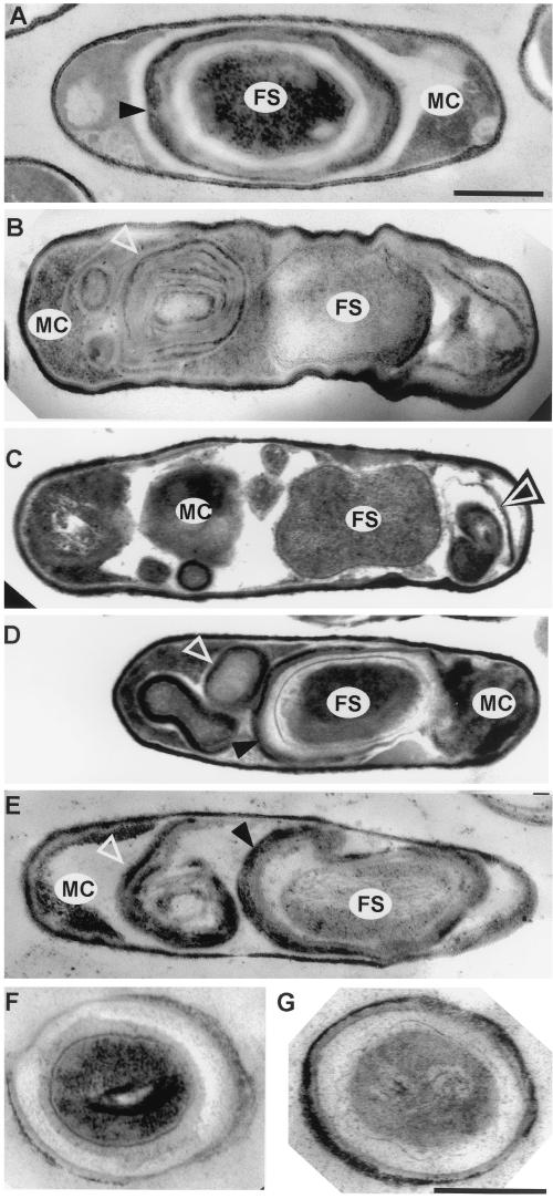
Electron microscopic examination of wild-type and spoIVA mutant sporulating cells. Thin-section electron micrographs of cells after 10 h of sporulation are shown. (A) PY79 (wild type); (B) AD18 (spoIVAΔ::neo); (C) FC336 (L393P); (D and E) FC329 (I400P, E418V, I457R); (F and G) FC297 (L59P) rehydrated spores. FS, forespore; MC, mother cell. Open triangles indicate swirls of coat; arrowheads indicate forespore-associated coat. Bars in panels A (for panels A to E) and G (for panels F and G), 500 nm.

