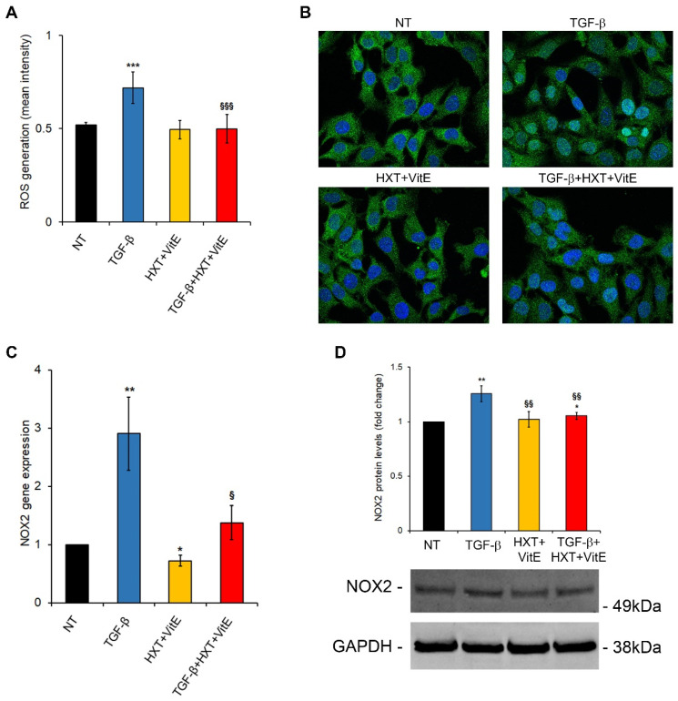
Figure 3.
HXT + VitE improves TGF-β-pro-fibrogenic phenotype by attenuating the SMAD/NOX2 pathway. (A) ROS levels assessed by using CM-H2DCFDA in LX-2 cells NT, treated with HXT + VitE, or treated with TGF-β alone or with HXT + VitE for 24 h. Data are expressed as the mean ± SD of at least five independent experiments. *** p < 0.001 vs. NT; §§§ p < 0.001 vs. TGF-β. (B) Representative images of SMAD2/3 (green) cellular localization by IF in LX-2 cells NT, treated with HXT + VitE, or treated with TGF-β alone or with HXT + VitE for 3 h. Nuclei were stained by Hoechst (blue). Magnification 60×. (C) NOX2 gene expression by qRT-PCR in LX-2 cells NT, treated with HXT + VitE, or treated with TGF-β alone or with HXT + VitE for 24 h. Data are expressed as the mean ± SD of three independent experiments. * p < 0.05 and ** p < 0.01 vs. NT; § p < 0.05 vs. TGF-β. (D) Quantitative analysis and representative immunoblot of NOX2 protein expression by WB in LX-2 cells NT, treated with HXT + VitE, or treated with TGF-β alone or with HXT + VitE for 24 h. GAPDH protein levels were used as loading control. Data are expressed as the mean ± SD of three independent experiments. * p < 0.05, ** p < 0.01 vs. NT; §§ p < 0.01 vs. TGF-β.

