Figure 4.
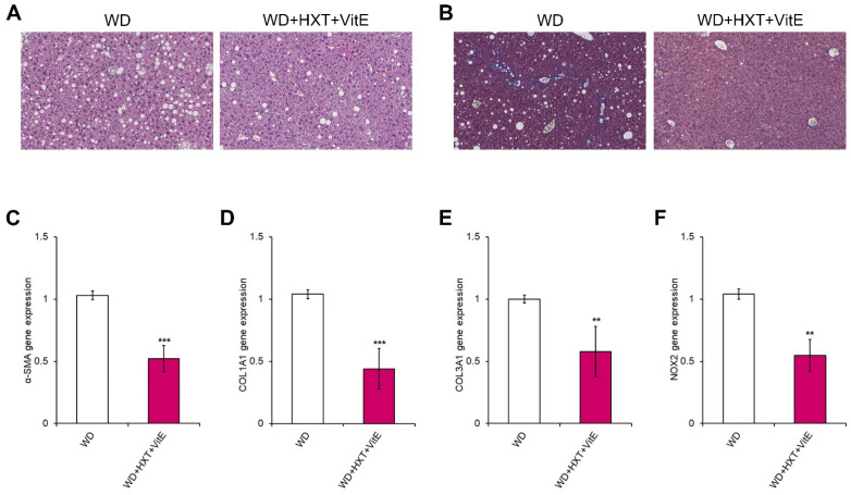
HXT + VitE improves WD-induced liver fibrosis in mice. Representative images of H&E (A) and Mason’s trichrome (B) staining in liver sections of WD and WD + HXT + VitE mice (20× magnification). Hepatic gene expression by qRT-PCR of α-SMA (C), COL1A1 (D), COL3A1 (E), and NOX2 (F) in WD and WD + HXT + VitE mice. Data are expressed as the mean ± SD of at least three independent experiments. ** p < 0.01 and *** p < 0.001 vs. WD.

