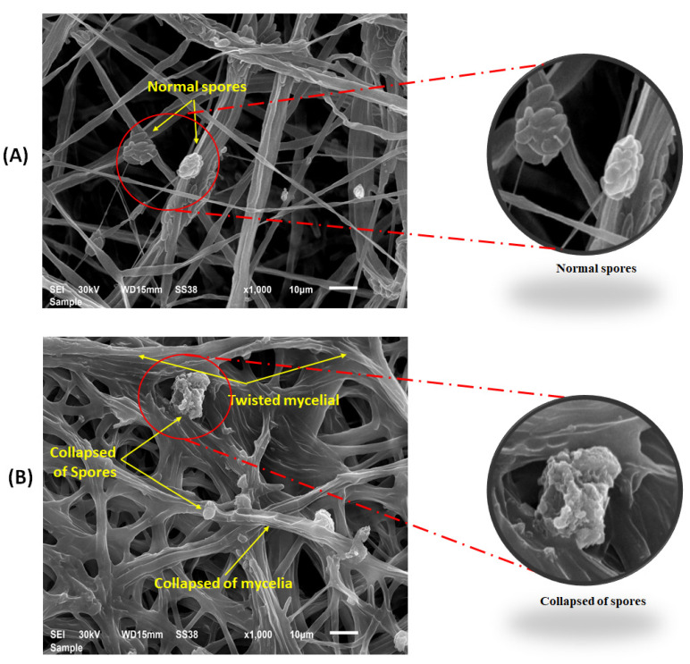
Figure 6.
Scanning electron microscope observations of spores and mycelia of F. solani taken from growth medium on potato dextrose agar, with two sizes of nano copper showing. (A): Untreated control with normal spores and mycelium (yellow arrows). (B): Treated with Cu2ONPs, showing collapsed mycelia and spores (yellow arrows).

