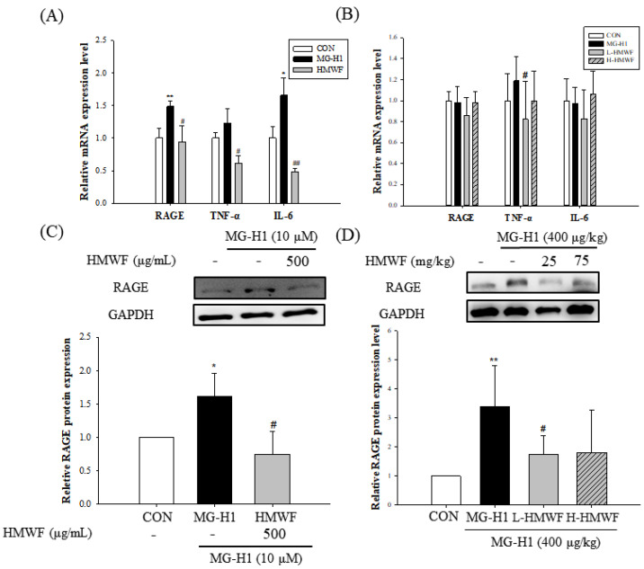
Figure 6.
Effect of HMWF on RAGE-induced intestinal inflammation. Caco-2 cell monolayers were incubated with 500 μg/mL HMWF and 10 μM MG-H1 for 24 h. Mice were treated with HMWF (25 or 75 mg/kg b.w.) orally and MG-H1 (400 μg/kg b.w.) intravenously once a day for 4 weeks. (A) The mRNA expression of RAGE, TNF-α, and IL-6 in Caco-2 monolayers and (B) mouse colon tissue. (C) Protein expression of RAGE in Caco-2 monolayers and (D) mouse colon tissue. The bar graph of the relative intensities of western blotting bands. All values are presented as the mean ± SD. The data were analyzed by Dunnett’s t-test. * p < 0.05, ** p < 0.01 versus the control group, # p < 0.05, ## p < 0.01 versus the MG-H1-treated group.

