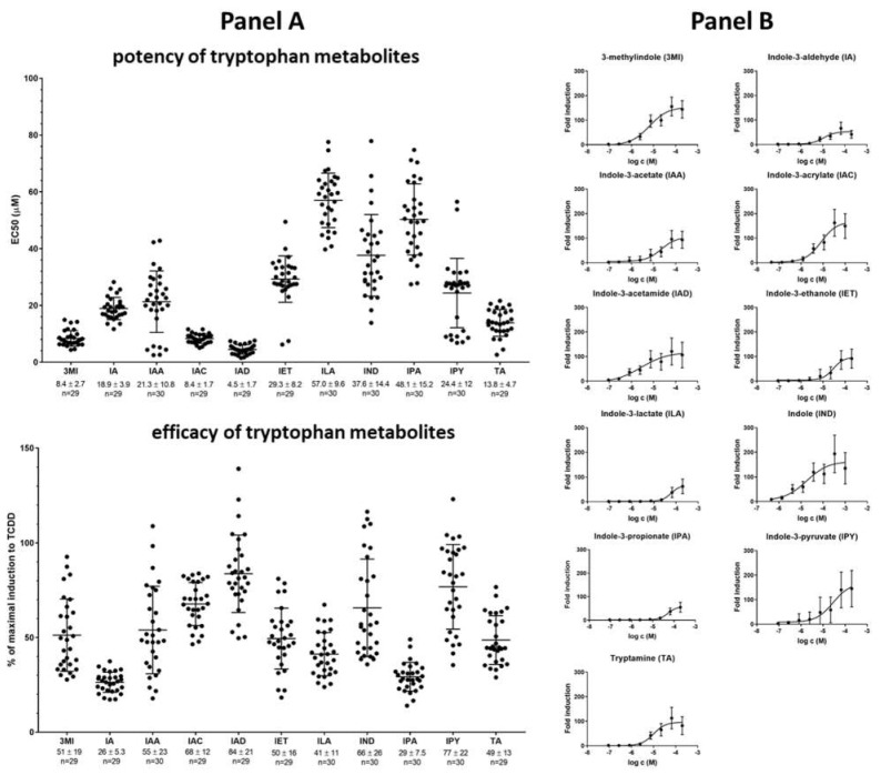
Figure 2.
High-throughput screening of AHR transcriptional activity by tryptophan microbial catabolites (MICT). AZ-AHR cells were incubated for 4 h with vehicle (DMSO; 0.1% v/v), TCDD (5 nM), or increasing concentrations of the tested compounds. Following the treatments, cells were lysed, and luciferase activity was measured. Panel (A): The column scatter plot represents the values of the half-maximal effective concentrations (potency; EC50) and relative efficacies (a ratio of luciferase activity by MICT in highest concentration/luciferase activity by TCDD). Panel (B): Dose–response assessment of AHR-dependent activity. Data are expressed as the fold induction of luciferase activity over control cells and are the mean ± SD from approximately 30 consecutive cell passages. The fold induction of TCDD was 200 ± 105 (n = 165). All values are statistically significant (p < 0.01) in comparison to vehicle-treated cells.

