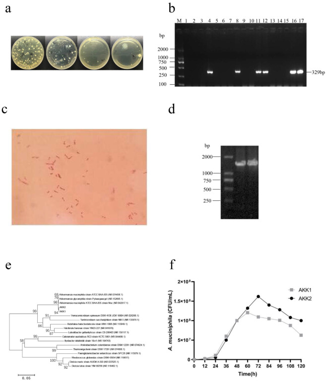
Figure 1.

A. muciniphila strains were isolated from mouse feces. (a) The colony morphology on vancomycin solid medium. (b) The PCR gel electrophoresis of suspected A. muciniphila strains. M: DL2000 DNA Marker; 1–17: suspected isolates. (c) Gram staining of strains (1000×). (d) PCR products of isolates AKK1 and AKK2 on agarose gel. M, DL2000 DNA Marker; 1–2: Suspected isolates. (e) Phylogenetic analysis of strains by maximum likelihood method and the general time reversible model. (f) growth curve of A. muciniphila AKK1 and AKK2.
