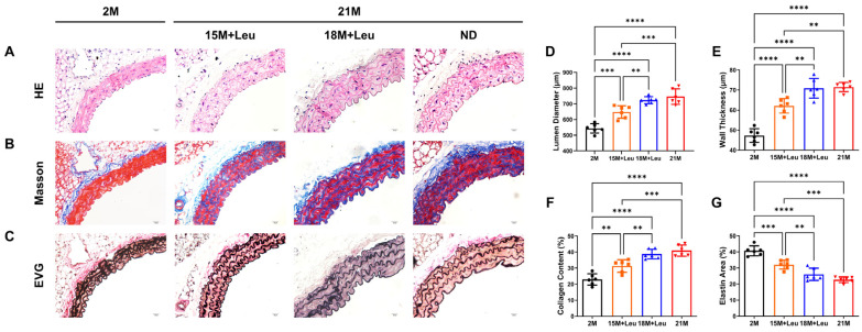
Figure 2.
Leucine supplementation from 15M improved aging-induced vascular remodeling. Representative images of H&E (A), Masson’s (B), and van Gieson’s (C) staining show the aortic structure of different groups at low and high magnifications. Lumen diameter and wall thickness of each group were measured (D,E). The percentage of collagen deposition area is shown in (F). Quantification of elastin area is shown in (G). Statistical analysis was performed using one-way ANOVA; data are presented as mean ± SEM, n = 6/group. ** p < 0.01, *** p < 0.001, and **** p < 0.0001. ND: normal diet.

