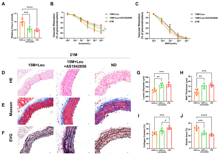
Figure 5.
Inhibition of Foxo1 activity reversed the protective effects of leucine supplementation on vascular dysfunction and remodeling. Quantification of Foxo1 activation (A). Dose–response curves for acetylcholine-mediated endothelium-dependent relaxation (B) and SNP-mediated endothelium-independent relaxation (C). Representative images of H&E, Masson’s, and van Gieson’s in aortas from mice at 2M and 21M, and 21M mice with leucine supplementation from 15M, with or without Foxo1 inhibitor AS1842856 (D–F). Quantitative analysis is shown in (G–J). Statistical analysis of vascular relaxation curves was performed with two-way ANOVA, * p < 0.05 15M + Leu + AS1842856 vs. 2M group, † p < 0.05 21M vs. 2M group, †† p < 0.01 21M vs. 2M group. Histological statistical analysis was performed with one-way ANOVA, * p < 0.05, ** p < 0.01, *** p < 0.001, and **** p < 0.0001. All data are presented as mean ± SEM, n = 6/group. ND: normal diet.

