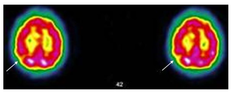
Figure 5.
Case MGA005; SPECT-CT (ECD Tc-99m) of 25/05/21; The image shows two brain sections of the same patient. The red areas indicate a good tracer fixation and thus a good perfusion. Yellow patches (see arrows) appearing in red areas indicate hypofixation and thus ischemia Protocol; “heterogeneous tracer fixation with clearer hypofixations left frontal, left parietal, right parietal. No preservation of the sensory-motor cortices. The fixation in front of the grey nuclei and the cerebellum is correct. Presence of periventricular hypocaptation. Conclusion: Evidence of heterogeneous tracer fixation and periventricular hypocaptation compatible with vascular-type cerebral damage.” (Images and protocol; Drs Bouazza & Mahy, Vésale Hospital, ISPPC, Belgium).

