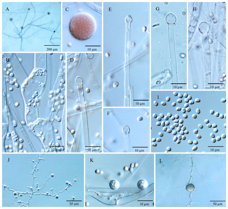
Figure 3.
Umbelopsis dura. (A) The main branching pattern of sporangiophore. (B) Branch point of sporangiophore. (C) Sporangium at tip of sporangiophore. (D–H) Various shapes of collars and columellae at sporangiophore tips after the sporangia have been dissolved. (I) Sporangiospores. (J,K) Micro-chlamydospores. (L) Macro-chlamydospore.

