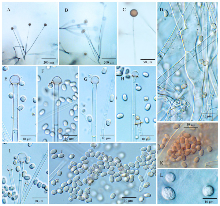
Figure 4.
Umbelopsis macrospora. (A,B) Branched sporangiophores. (C) Sporangium at tip of sporangiophore. (D) Branch point of sporangiophore. (E–I) Various shapes of collars and columellae at sporangiophore tips after the sporangia have been dissolved. (J,K) Sporangiospores. (L) Micro-chlamydospores.

