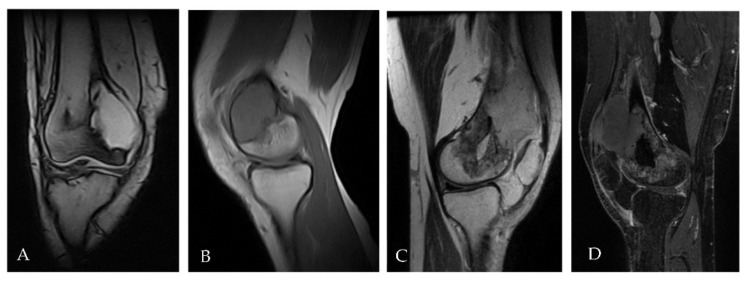
Figure 3.
(A,B) MRI performed at time of initial diagnosis showed a focal eccentric lesion located in the meta-epiphyseal of distal right femur without interruption of cortical bone and/or involvement of neighbouring soft tissue. (C,D) MRI performed after surgery with curettage six months later showed the presence of solid heterogeneous high signal tissue in T2 (3C) enhancing post contrast (3D) with large extension in the soft tissue compatible with relapse.

