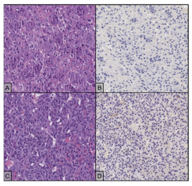
Figure 4.
(A) The lesion is composed by multinucleated and mononuclear cells (H&E stain, 20×); the giant cells have hyperchromatic, homogenous nuclei, while the mononuclear cells tend to present slightly larger nuclei with finely dispersed chromatin and evident nucleolus. (B) Both cell populations test negative for H3F3A immunostaining (20×). (C) Recurrent disease shows proliferation of atypical, round cells, with no evidence of giant cells. (H&E stain, 20×). (D) H3F3A immunostaining is negative in the neoplastic cells (20×).

