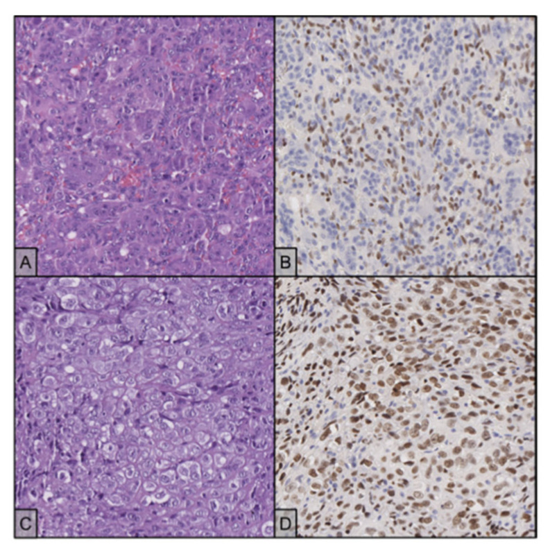
Figure 8.
(A) The primary disease shows typical histology with osteoclast-like giant cells between numerous mononuclear cells (H&E stain, 20×); the two populations showed remarkably similar nuclear features, delicate nucleoli and finely dispersed chromatin; the interposed stroma appeared haemorrhagic. (B) Mononuclear cells react with an antibody against H3F3A, while the giant cells are negative (20×). (C) Recurrent disease shows different morphological features and no evidence of giant cells (H&E stain, 20×). (D) Neoplastic cells maintain positivity for H3F3A immunostaining (20×).

