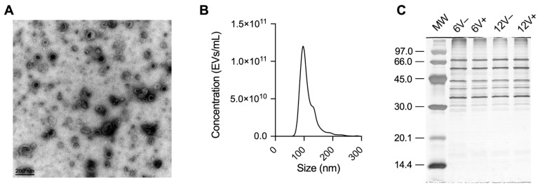
Figure 1.
S. aureus HG003 EV characterization. (A) Cup-shaped EVs by transmission electron microscopy (TEM). (B) Monodisperse profile revealed by nanoparticle tracking analysis (NTA). (C) Protein profile of EV samples resolved in 12% SDS-PAGE. Molecular weight (MW) standards are indicated in kDa. Early- and late-stationary growth phases (6 and 12 h, respectively) in the absence (V−) or presence (V+) of vancomycin.

