Figure 2.
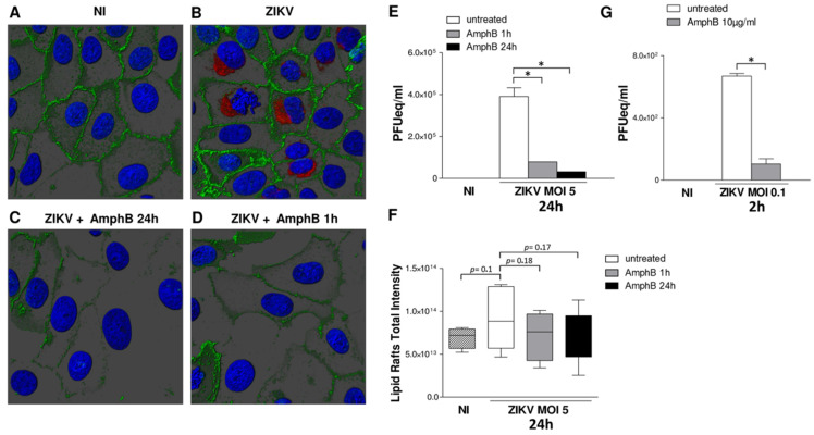
ZIKV exploits lipid rafts to enter and infect Vero cells. Vero cells were adsorbed with ZIKV at MOI 5 for 1 h at 37 °C in medium +/− AmphB (10 µg/mL). Unbound virus was removed, and infected cells were cultured in fresh medium +/− AmphB for 24 h or the drug was added only during 1 h of virus adsorption. At 24 h post-infection, cells were stained with the lipid raft marker CTB (green), fixed and stained with anti-pan-flavivirus antibody (ZIKV) followed by donkey anti-mouse Alexa Fluor 594 antibody (red). Nuclei were stained with DAPI (blue). 3D images were visualized and analysed by Imaris v8.1.2 software. NI: non-infected. (A,B) As compared to NI cells, ZIKV infection induced a lipid raft reorganization on cell surface. (C) Treatment with AmphB for 24 h disrupted lipid raft architecture and inhibited cell infection. (D) Treatment with AmphB only along 1 h of ZIKV adsorption disrupted lipid raft architecture and impaired the first steps of viral entry. Images are shown from one representative experiment out of three. (E) Viral titre was determined by qRT-PCR and expressed as PFU equivalents/mL (PFUeq/mL). Both drug treatments significantly inhibited viral replication. (F) Quantification of lipid rafts total intensity by 3D image analysis. The 3D surface enclosing the lipid raft signal was generated using the same parameters for all the images. The Sum Fluorescent Intensity inside the volume was measured and results summarized in the graph. ZIKV infection increased the lipid rafts fluorescence intensity, whereas both drug treatment decreased this value. (G) Treatment of AmphB during the first two hours of ZIKV infection (MOI 5) reduced viral entry as measured by titration of viral RNA extracted from infected cells and expressed as PFU equivalents/mL (PFUeq/mL). Results are expressed as mean ± SD of three independent experiments performed. Significance was determined by the nonparametric Mann–Whitney U test using GraphPad Prism. * p value < 0.05 was considered statistically significant.

