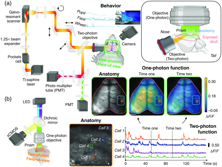
Fig. 1.
Simultaneous one-photon (wide-field) imaging and two-photon imaging. (a) A schematic of the light path of the two-photon imaging microscope (left). The FOV of the one-photon imaging setup is indicated by a green box. Behavioral data are collected using an auxiliary camera (middle). A schematic of the surgery—skull thinning, cranial window, and placement of a small prism—is shown in the in-lay (upper right). (b) A schematic of the light path of the one-photon imaging microscope (left). The FOV of the multiphoton imaging setup is indicated by the orange box. Example data are shown (right) (adapted with permission from Refs. 43 and 44).

