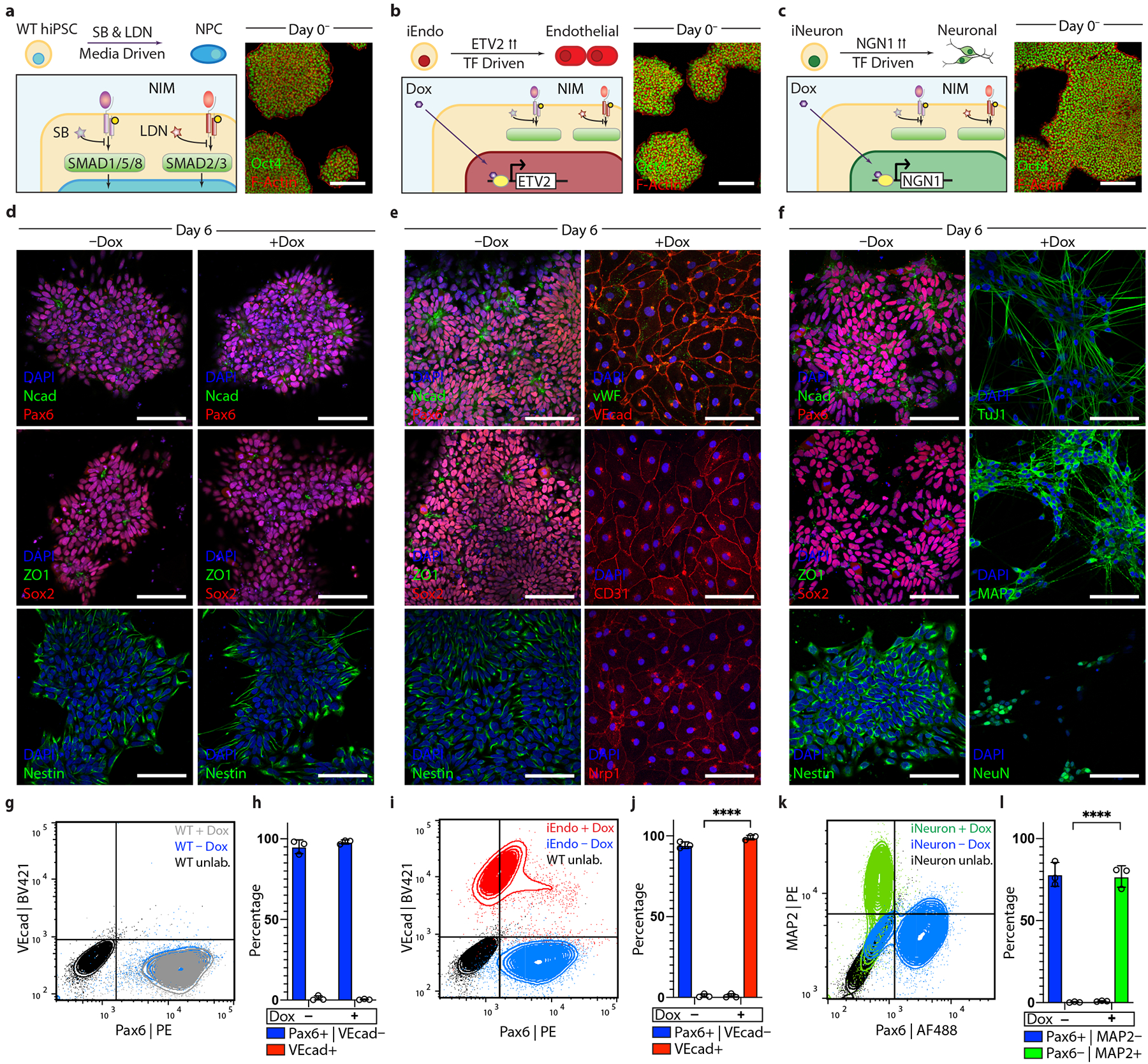
Fig. 2 |. Programmable differentiation of pluripotent stem cells via OiD under identical media conditions.

a, Left: schematic detailing WT hiPSC differentiation into neural stem cells in NIM. right: immunostaining of Oct4 and F-actin of WT colonies on day 0. b, Left: schematic detailing iEndo differentiation through doxycycline-induced ETV2 isoform-2 overexpression. right: immunostaining of Oct4 and F-actin of iEndo colonies on day 0. c, Left: schematic detailing iNeuron differentiation through doxycycline-induced NGN1 overexpression in culture. right: immunostaining of Oct4 and F-actin of iNeuron colonies on day 0. d, WT hiPSCs cultured in NIM for 6 d without (left column) or with (right column) doxycycline. Immunostaining of NCAD and PAX6, ZO1 and SOX2, and nestin. e, iEndo cells cultured in NIM for 6 d without (left column) or with (right column) doxycycline. Left: immunostaining of NCAD and PAX6, ZO1 and SOX2, and nestin. right: immunostaining of VWF and VEcad, CD31 and NrP1. f, iNeuron cells cultured in NIM for 6 d without (left column) or with (right column) doxycycline. Left: immunostaining of NCAD and PAX6, ZO1 and SOX2, and nestin. right: immunostaining of Tuj1, MAP2 and NeuN. g, Flow cytometry plots quantifying WT hiPSC differentiation to PAX6+ neural stem cells by 6 d with (grey) and without (blue) doxycycline. BV, Brilliant Violet 421; PE, phycoerythrin. h, Quantification of populations, mean ± s.e.m., n = 3 biological replicates. i, Flow cytometry plots indicating that iEndos differentiate into VEcad+ endothelium in the presence of doxycycline (red) or into PAX6+ neural stem cells in its absence (blue). j, Quantification of populations, mean ± s.e.m., n = 3 biological replicates, **** P = 7.06 × 10⁻⁷, unpaired two-tailed t-test. k, Flow cytometry plots indicating that iNeurons differentiate into MAP2+ neurons in the presence of doxycycline (green) or into PAX6+ neural stem cells in its absence (blue). AF488, Alexa Fluor 488. l, quantification of populations, mean ± s.e.m., n = 3 biological replicates, ****P = 3.53 × 10⁻⁵, unpaired two-tailed t-test. Scale bars: 100 μm in d–f; 50 μm in a–c.
