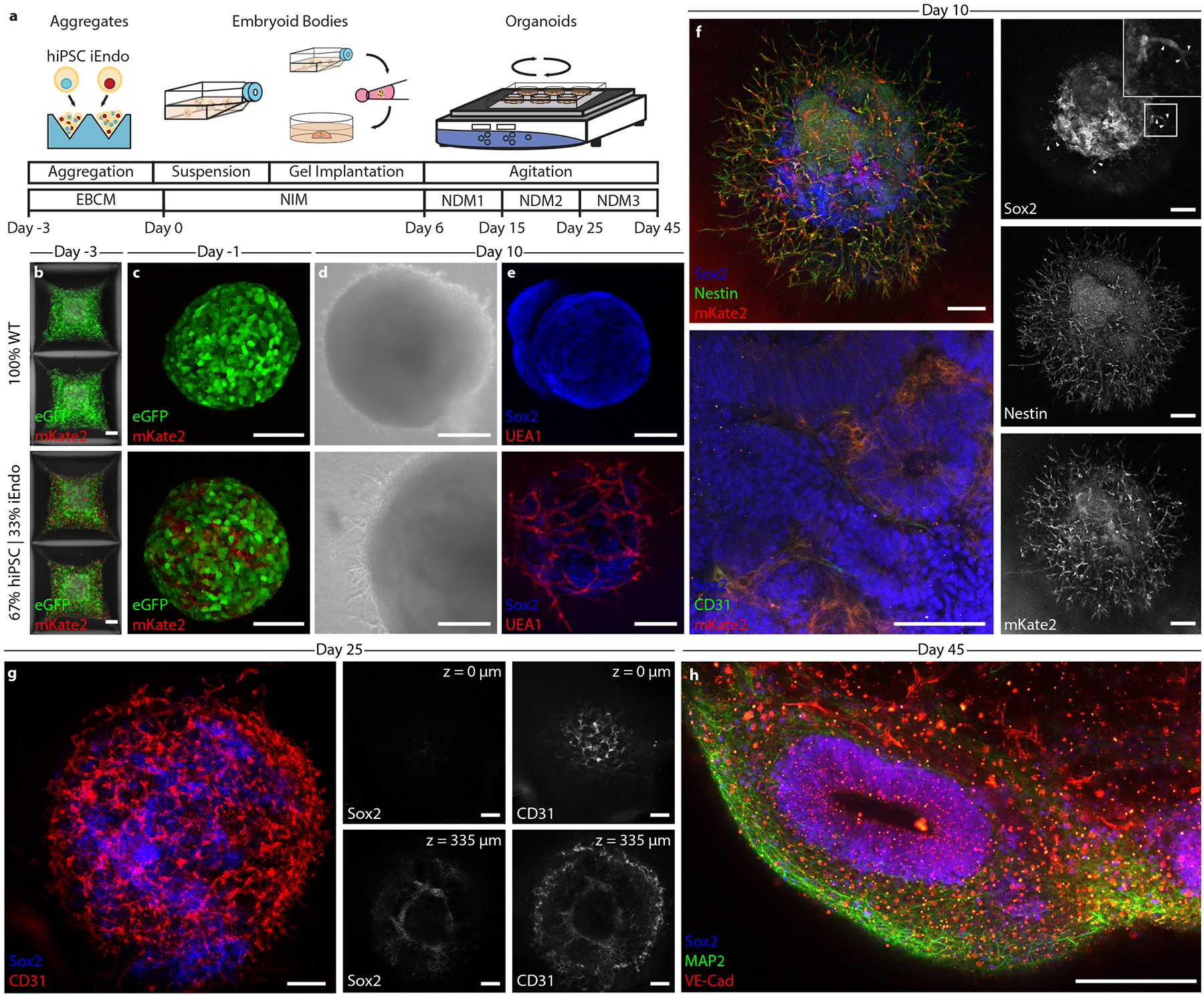
Fig. 3 |. Programmable vascularization of cortical organOiDs.

a, Schematic of the vascularized cortical organoid protocol. b, Fluorescent images of hiPSCs in microwells 3 d before suspension culture. Top: 100% WT-eGFP hiPSCs. Bottom: 67% WT-eGFP and 33% iEndo-mKate2 randomly pooled hiPSCs. c, Fluorescent images of resulting EBs. Top: 100% WT-eGFP EBs. Bottom: 67% WT-eGFP and 33% iEndo-mKate2 randomly pooled EBs. d, Brightfield images of organoids derived from EBs cultured for 10 d. Top: 100% WT-eGFP organoids. Bottom: 67% WT-eGFP and 33% iEndo-mKate2 pooled organoids. e, Immunostaining of SOX2 and UEA1 labelling of organoids cultured for 10 d. Top: 100% WT organoids. Bottom: 67% WT and 33% iEndo pooled organOIDs. f, Left, top: confocal maximum intensity z-projections obtained from immunostaining of SOX2 and nestin of 67% WT and 33% iEndo-mKate2 pooled organOIDs cultured for 10 d; right column: individual channels for SOX2, nestin and mKate2. Arrowheads indicate SOX2+ positive cells that co-localize with the vasculature. Left, bottom: immunostaining of SOX2 and CD31 with iEndo-mKate2 of 67% WT and 33% iEndo-mKate2 pooled organOIDs cultured for 10 d. g, Left: maximum intensity z-projection with immunostaining of SOX2 and CD31 of 67% WT and 33% iEndo pooled organOIDs cultured for 25 d. right: optical slices of SOX2 and CD31 channels at depths of z = 0 μm (top row) and z = 335 μm (bottom row). h, Maximum intensity z-projection with immunostaining of SOX2, MAP2 and VEcad of 67% WT and 33% iEndo pooled organOIDs cultured for 45 d. Scale bars: 200 μm in d, f (top), g and h; 100 μm in b, c, f (bottom) and e.
