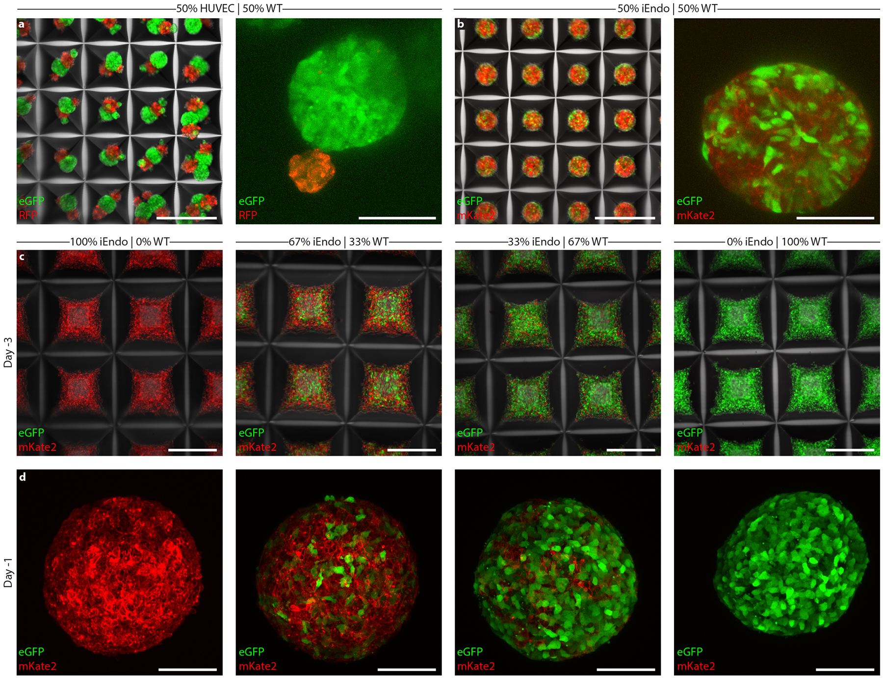
Extended Data Fig. 1 |. Pooled hiPSCs enable the formation of cohesive embryoid bodies with tailorable cellular composition.

a, Left, 50% WT-eGFP and 50% rFP HUVEC EBs in microwell arrays cultured for 1 day. right, 50% WT-eGFP and 50% rFP HUVEC EBs cultured for 3 days. b, Left, 50% WT-eGFP and 50% iEndo-mKate2 EBs in microwell arrays cultured for 1 day. right, WT-eGFP and 50% iEndo-mKate2 EBs cultured for 3 days. c, Different seeded proportions of WT-eGFP and iEndo-mKate2 hiPSC aggregates in microwell arrays 3 days before suspension culture. Left, 100% iEndo-mKate2. Middle-left, 67% iEndo-mKate2 and 33% WT-eGFP. Middle-right 33% iEndo-mKate2 and 67% WT-eGFP. right, 100% WT-eGFP. d, WT-eGFP and iEndo-mKate2 EBs 1 day before suspension culture. Left, 100% iEndo-mKate2. Middle-left, 67% iEndo-mKate2 and 33% WT-eGFP. Middle-right 33% iEndo-mKate2 and 67% WT-eGFP. right, 100% WT-eGFP. Scale bars: 500 μm in a (left), b (left), and c; 100 μm in a (right), b (right), and d.
