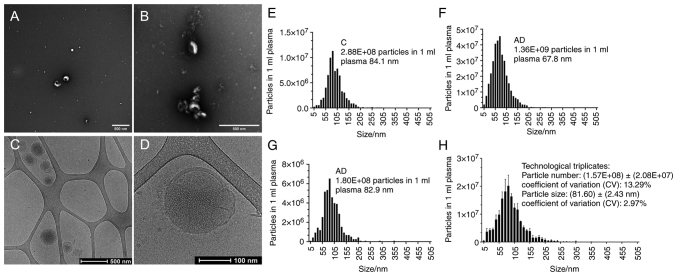
Figure 2.
EV morphology and size distribution. (A and B) Eluted EVs by EXÖBead® isolation in transmission electron microscopy. (C and D) Eluted EVs by EXÖBead® isolation in cryo-electron microscopy. (E-G) Particle size distribution of eluted EVs by EXÖBead® isolation from three individual donors, measured with Zetaview®. (H) Particle size distribution of eluted EVs by EXÖBead® isolation from the same donor with three independent experiments, measured with Zetaview®. EV, extracellular vesicle.

