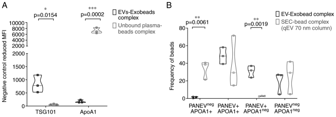
Figure 4.
Bead-based flow cytometry analysis of EV intracellular marker and non-EV marker. (A) Intracellular EVs markers (TSG101) and non-EV markers (ApoA1) of plasma EVs-EXÖBead® complexes and unbound plasma fraction magnetic bead complexes (n=3) are shown as reduced geometric mean fluorescence intensity in the negative control. (B) PanEV+/Neg and ApoA1+/Neg populations of the plasma EVs-EXÖBead® complex and SEC (Izon qEVoriginal 70) plasma EVs-magnetic beads complex are expressed as percentages by gating with FlowJo™. Significance was calculated using an unpaired t-test. EV, extracellular vesicle.

