. 2022 Aug 8;11:e77035. doi: 10.7554/eLife.77035

Figure 1. Vrl1 is a predicted PX-BAR protein that interacts with conserved machinery at the endosome.

(A) Schematic of Vrl1 and VARP domain architecture. (B) ColabFold predicts the Vrl1 C-terminus has a SNX-BAR-like PX and BAR domain fold. (C) Vrl1-Envy colocalizes with Did2-mRuby2-labeled endosomes, but not with the Sec7-dsRed Golgi marker. (D) Quantification of colocalization as the percentage of Vrl1 puncta overlapping RFP puncta in C. Two-tailed equal variance t test; n=3, cells/strain/replicate ≥1395; ****=p < 0.0001. (E) Schematic of DHFR proximity screen methodology. (F) Z-score distribution of the ratio of colony areas from genome-wide DHFR screens of full-length and truncated Vrl1 baits that localize to the endosome and cytosol, respectively. (G) Gene Ontology (GO) functional enrichment analysis of Vrl1 DHFR interactors (Z-score >2; http://geneontology.org). GO terms of the most specific hierarchical subclass with a fold enrichment value >25 are presented as the negative base 10 log of the associated p-value from a Bonferroni-corrected binomial test of significance. Scale bars, 2 µm. Error bars report standard error of the mean (SEM). Enrich., enrichment. aa, amino acids.

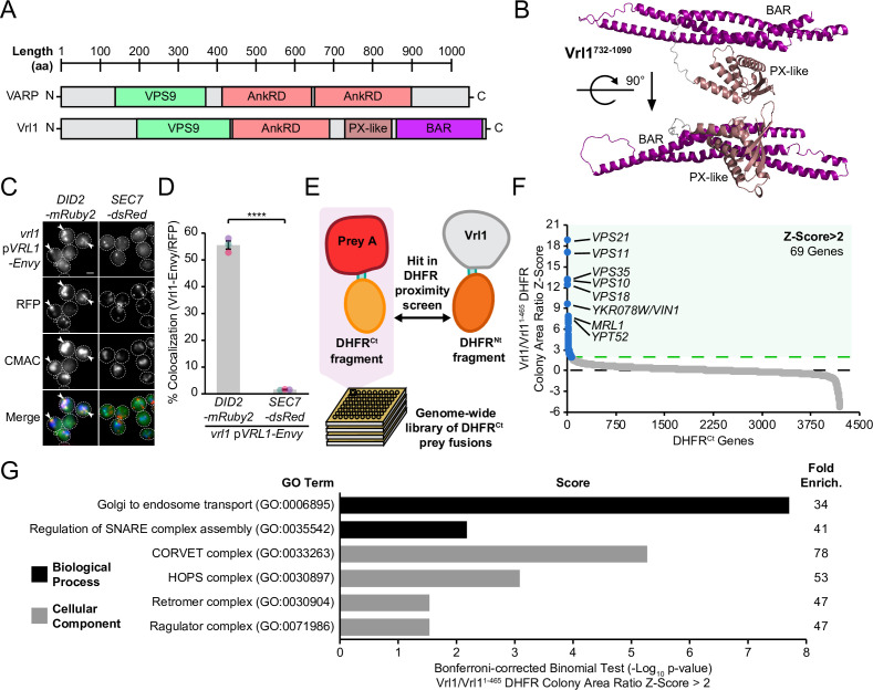
Figure 1—source data 1. Data associated with Figure 1D.
Figure 1—source data 2. Data associated with Figure 1G.

Figure 1.

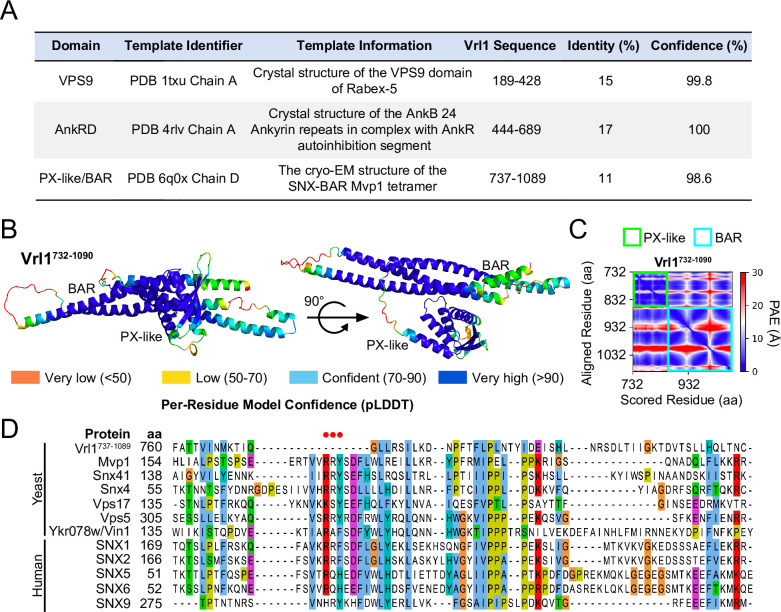
Figure 1—figure supplement 1. The Vrl1 PX-like domain is missing key PI3P-binding residues.

Figure 1—figure supplement 1.

(A) Results from Phyre2 analysis of Vrl1 sequences (Intensive mode, http://www.sbg.bio.ic.ac.uk/phyre2). (B) ColabFold-predicted Vrl1 C-terminus with the predicted local-difference distance test (pLDDT; Jumper et al., 2021; Mirdita et al., 2022) scores mapped to each residue. (C) ColabFold-generated predicted alignment error (PAE; Jumper et al., 2021; Mirdita et al., 2022) plot for the Vrl1 C-terminus demonstrates a high confidence folding prediction. (D) Sequence alignment of Vrl1 C-terminus (aa 737–1089) with yeast and human PX domain-containing proteins. The canonical PI3P-binding ‘RRY’ motif is absent in Vrl1 (highlighted with red dots above alignment). aa, amino acids.
Figure 1—figure supplement 2. The Vrl1 N-terminus and VPS9 domain are not sufficient to localize to puncta.

Figure 1—figure supplement 2.

The Vrl1 N-terminus (aa 1–465) does not localize to puncta when expressed from the endogenous VRL1 promoter (VRL1pr) or the strong ADH1pr and instead accumulates in the cytosol. Insets are scaled to match other images in the same channel (see materials and methods for details). Scale bars, 2 µm. WT, wild type.