. 2022 Aug 8;11:e77035. doi: 10.7554/eLife.77035

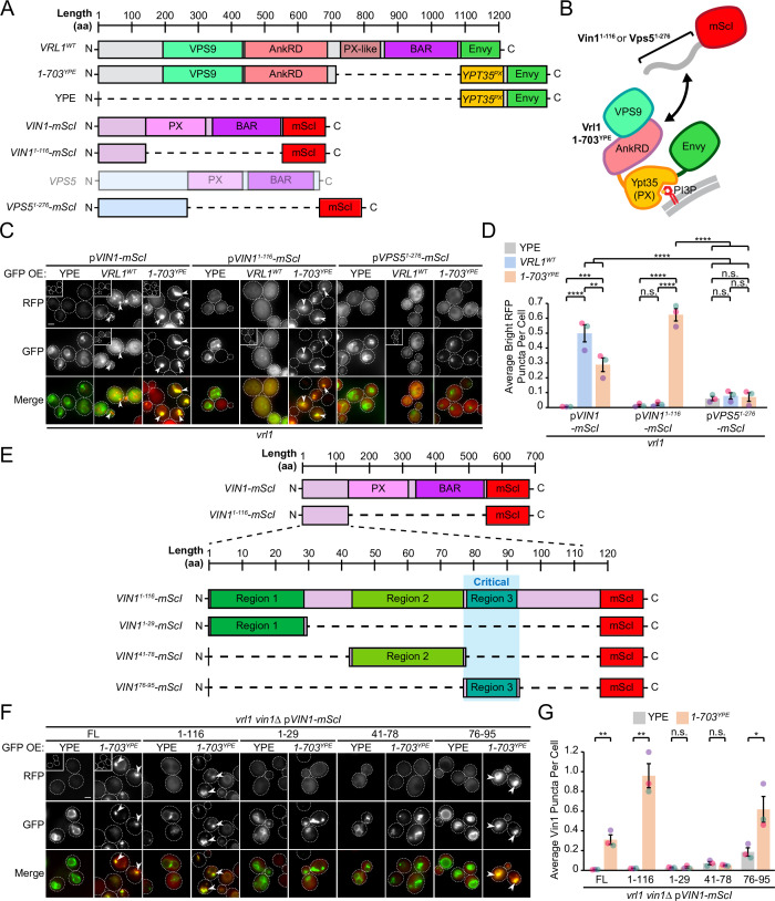
Figure 4. The Vrl1 AnkRD recognizes a small region of the Vin1 N-terminus.

(A) Schematic of constructs used in C, D. Full-length Vps5 was not tested but is shown for comparison. (B) Diagram of chimeric Vrl1 recruitment assay used to test for interactions with the unstructured N-terminus of either Vps5 (Vps51-276) or Vin1 (Vin11-116). (C) The AnkRD-containing Vrl1(1-703)YPE chimera recruits the N-terminus of Vin1, but not Vps5. Insets are scaled to match other images in the same channel (see Materials and methods for details). (D) Quantification of RFP puncta per cell in C. One-way ANOVA with Tukey’s multiple comparison test; n=3, cells/strain/replicate ≥902; not significant, n.s.=p > 0.05, **=p < 0.01, ***=p < 0.001, ****=p < 0.0001. (E) Schematic of Vin1 N-terminal fragments used to map the Vrl1 recruitment site. (F) The AnkRD-containing Vrl1(1-703)YPE chimera recruits a small fragment of the Vin1 N-terminus. Insets are scaled to match other images in the same channel. (G) Quantification of Vin1-mScI puncta per cell in F. Two-tailed equal variance t tests; n=3, cells/strain/replicate ≥294; not significant, n.s.=p > 0.05, *=p < 0.05, **=p < 0.01. Scale bars, 2 µm. Error bars report SEM. OE, over-expressed. FL, full-length. WT, wild type. YPE, Ypt35(PX)-Envy.

Figure 4—source data 1. Data associated with Figure 4D.
Figure 4—source data 2. Data associated with Figure 4G.

Figure 4.

Figure 4—figure supplement 1. The N-terminal regions of Vin1 and Vps5 are predicted to be disordered.
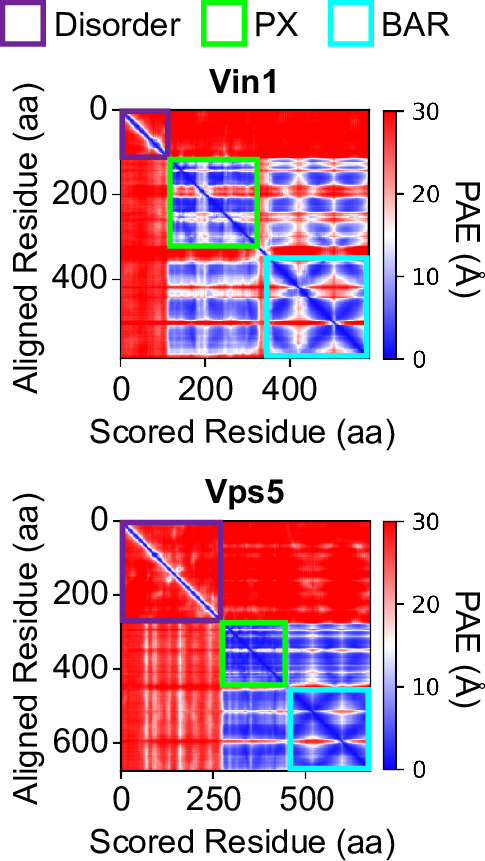
Figure 4—figure supplement 1.

A lack of off-diagonal signal in the ColabFold-generated predicted alignment error (PAE; Jumper et al., 2021; Mirdita et al., 2022) plots of Vin1 and Vps5 indicates a shared lack of structure in the N-terminus of either protein. On-diagonal signal for the PX and BAR domains provides a point of comparison for how structured domains appear in prediction PAE plots. aa, amino acids.
Figure 4—figure supplement 2. The Vin1 PX-BAR region is indispensable for Vrl1 localization.

Figure 4—figure supplement 2.

(A) The Vin1 N-terminus is not sufficient to localize over-expressed Vrl1-Envy to membranes in a VIN1 deletion strain. The Vrl1(1-703)YPE chimera recruits the Vin1 N-terminal prey construct to puncta in strains lacking the chromosomal copy of VIN1. Insets are scaled to match other images in the same channel (see Materials and methods for details). (B) Quantification of RFP puncta per cell in A. One-way ANOVA with Tukey’s multiple comparison test; n=3, cells/strain/replicate ≥1068; not significant, n.s.=p > 0.05, **=p < 0.01, ***=p < 0.001, ****=p < 0.0001. Scale bars, 2 µm. Error bars report SEM. WT, wild type. YPE, Ypt35(PX)-Envy.
Figure 4—figure supplement 2—source data 1. Data associated with Figure 4—figure supplement 2B.
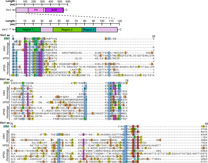
Figure 4—figure supplement 3. The Vin1 N-terminus has three conserved regions in fungal homologs.

Figure 4—figure supplement 3.

Sequence alignment of Vin1 and Vps5 orthologs collected using the Yeast Gene Order Browser (http://ygob.ucd.ie/). Three conserved regions were selected for expression as mScI-tagged fragments: Vin11-29 (Region 1), Vin141-78 (Region 2), and Vin176-95 (Region 3). aa, amino acids.