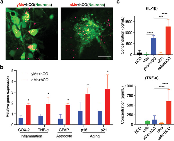
Figure 5.

Characterization of monocyte‐driven aging phenotypes. a) Confocal images of neuron morphology adjacent to infiltrated monocytes isolated from old donors (>60‐year‐old) into on‐chip cultured human cortical organoid (oMs+hCO) with monocytes isolated from young donors (20 to 30‐year‐old) into on‐chip cultured human cortical organoid (yMs+hCO). Scale bar: 20 µm. b) Comparison of proinflammation genes (COX‐2 and TNF‐α) and senescence genes (p16INK4a and p21CIP1) in monocytes within oMs+hCO and yMs+hCO cultures on day 29. 2−ΔΔCt calculated as “delta Ct” (∆∆Ct) of GAPDH and target gene normalized against on‐chip cultured human cortical organoid only on the same day, n = 3. c) Characterization of IL‐1β and TNF‐α concentrations in supernatant isolated from hCO, yMs, oMs, yMs+hCO, and oMs+hCO cultures, n = 6.
