
Figure 2.
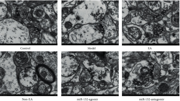
EA ameliorated damage to neuronal synaptic structures in REMSD rats. Transmission electron microscope detection of synapse in the CA1 regions of the hippocampus in each group. Scale bars, 500 nm. Presynaptic membrane (PM), post-synaptic membrane (PD) and synaptic vesicle (SV) showed in the representative pictures. n = 3 per group.