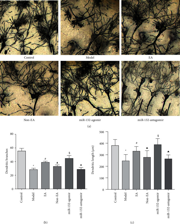
Figure 3.

EA attenuated lesions of neuronal dendritic structures in the hippocampus of REMSD Model Rats (a), representative pictures of Golgi staining in each group, scale bar, 100 μm (b, c), dendritic branches and dendritic length in each group were measured by Image J software. Data were presented as the mean ± s.d. One-way ANOVA was used for panels (a–c). ∗P < 0.05 versus control group; #P < 0.05 versus model group; ▲P < 0.05 versus EA group; and $ P < 0.05 versus non-EA group. n = 3 per group.
