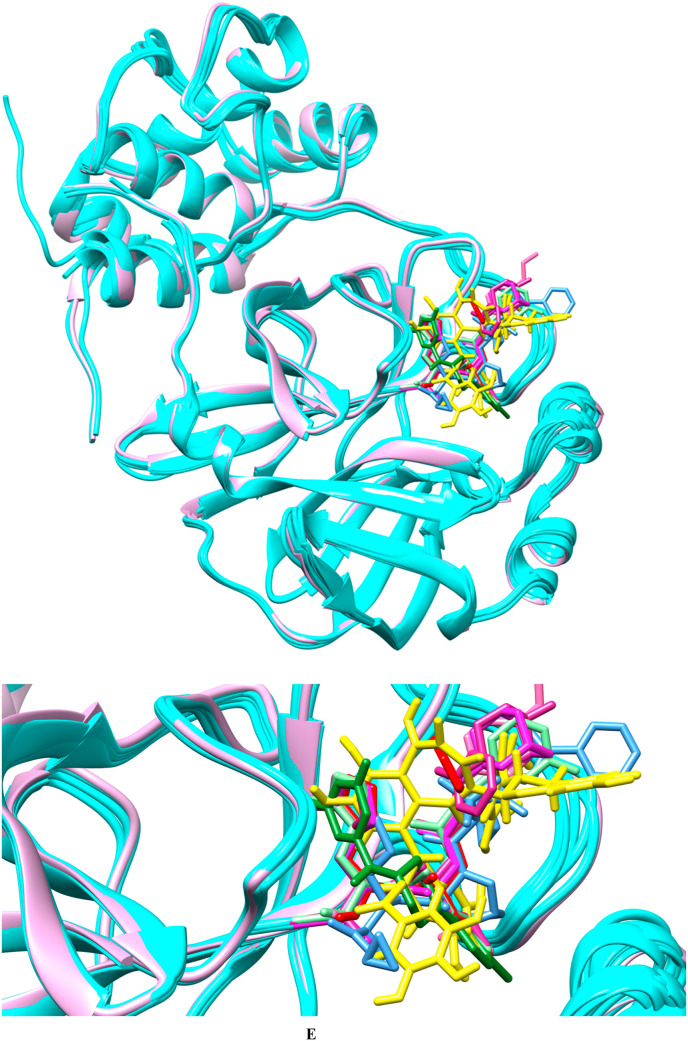
Fig. 1e.
Comparison of crystallographic poses of reference compounds with docked pose of TG. Superimposed protein crystal structure in cyan, whereas TG has been highlighted in yellow. The figure depicts noncovalent inhibitor YD1 in forest green, SV6 (telaprevir) in blue, YKS in violet-red, YLJ in light green, YNI in hot pink, and Y4V in green magenta, K36 (feline coronavirus inhibitor GC3760) in red. The upper panel represents the far view whereas lower panel represents the close view.

