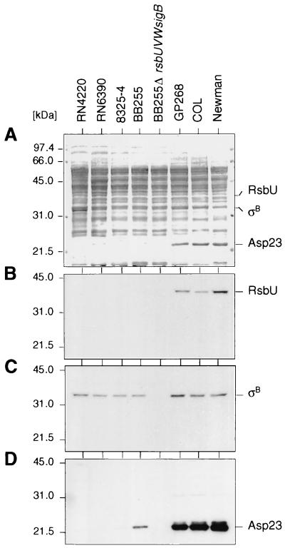
FIG. 3.
Western blot analyses of different S. aureus strains. Cytoplasmic protein fractions (10 μg/lane) of different S. aureus overnight cultures, grown in LB medium at 37°C and 200 rpm, were separated using SDS–10% PAGE and blotted onto nitrocellulose. The blotted proteins were either stained with amido black (A) or subjected to Western blot analyses using antigen-purified anti-RsbU antibodies (B), anti-SigB antibodies (C), or anti-Asp23 antibodies (D). The broad-range molecular size marker (Gibco-BRL) was used. Relevant protein signals are indicated.

