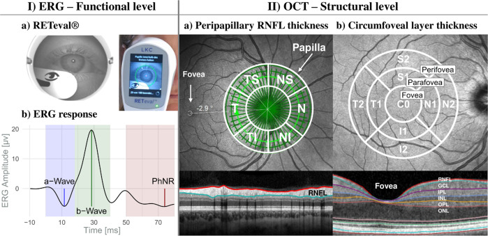
Fig. 1. Schematic overview of retinal examinations.
(I) ERG: a) Illustration of the RETeval® system during recording (right) and a picture from the device internal camera (left); b) Schematic retinal response comprising the a- and b-wave and the PhNR. (II) OCT: a) Peripapillary RNFL thickness from the circle scan with the Garway-Heath-grid; b) Circumfoveal scan for layer (macular, RNFL, GCL, IPL, INL, OPL, and ONL) thicknesses and volumes and the ETDRS grid (3 regions: fovea, parafovea, and perifovea). GCL ganglion cell layer, I inferior, INL inner nuclear layer, IPL inner plexifom layer, N nasal, ONL outer nuclear layer, OPL outer plexiform layer, RNFL retinal nerve fiber layer, S superior, T temporal.

