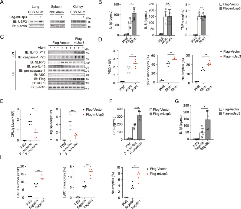
Fig. 6.
USP3 overexpression facilitates NLRP3, AIM2 and NLRC4 inflammasome activation in vivo. A Immunoblot analysis of the protein level of USP3 in various organs of WT- or USP3-overexpressing mice after i.p. Alum injection for 12 h. B ELISA analysis of IL-1β, TNF-α and IL-6 secretion from the peritoneal lavage fluid of WT- or USP3-overexpressing mice after i.p. Alum injection for 12 h. (mice: n = 5 for each group). C Immunoblot analysis of supernatants (SN) or cell lysates (CL) of lavage fluid from WT- or USP3-overexpressing mice after i.p. Alum injection for 12 h. (mice: n = 3 for each group). D Absolute numbers of PECs, percentages of neutrophils and Ly6C+ monocytes from WT- or USP3-overexpressing mice after i.p. Alum injection for 12 h. (mice: n = 5 for each group). E WT- or USP3-overexpressing mice were infected subcutaneously with 1.5 × 10⁵ CFU F. novicida, and the bacterial burden in the spleen and liver was measured on Day 2 after infection. (mice: n = 5 for each group). F ELISA analysis of IL-1β in sera from WT- or USP3-overexpressing mice infected subcutaneously with 1.5 × 10⁵ CFU F. novicida. G ELISA analysis of IL-1β in the BALF from WT- or USP3-overexpressing mice 12 h after intranasal instillation of isotonic saline or flagellin. H Absolute numbers of BALCs, percentages of neutrophils and Ly6C+ monocytes from WT- or USP3-overexpressing mice after isotonic saline or flagellin intranasal instillation for 12 h. Data are shown as the mean + S.D. in (B, F, G). Similar results were obtained in three independent experiments. *p < 0.05, **p < 0.01, ***p < 0.001; two-tailed Student’s t test

