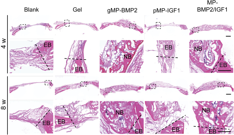
FIG. 4.
Representative images of H&E staining of cranial defect sites with native bone at 4 or 8 weeks after operation. The sections were made in coronal axial. Black dash frames indicate the locations of the images in higher magnification. Dash lines indicate the margin dividing EB and the newly formed tissue in cranial defect sites. Scale bar: 1 mm. EB, existing bone; H&E, Hematoxylin and Eosin; NB, new bone. Color images are available online.

