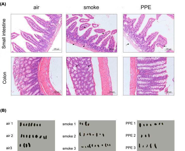
Figure 1. Mice model of emphysema displayed intestinal pathological changes.
(A) Comparison of H&E staining of the small intestine (upper, Scale bar = 100 μm) and colon (lower, Scale bar = 100 μm) from air-exposed control and emphysema model mice were performed (n=5 per group). (B) Effects of CS and PPE on the feces of C57BL/6 mice. Feces collected from air-exposed control and emphysema model mice under the normal diet for 1 h were represented (n=3 per group).

