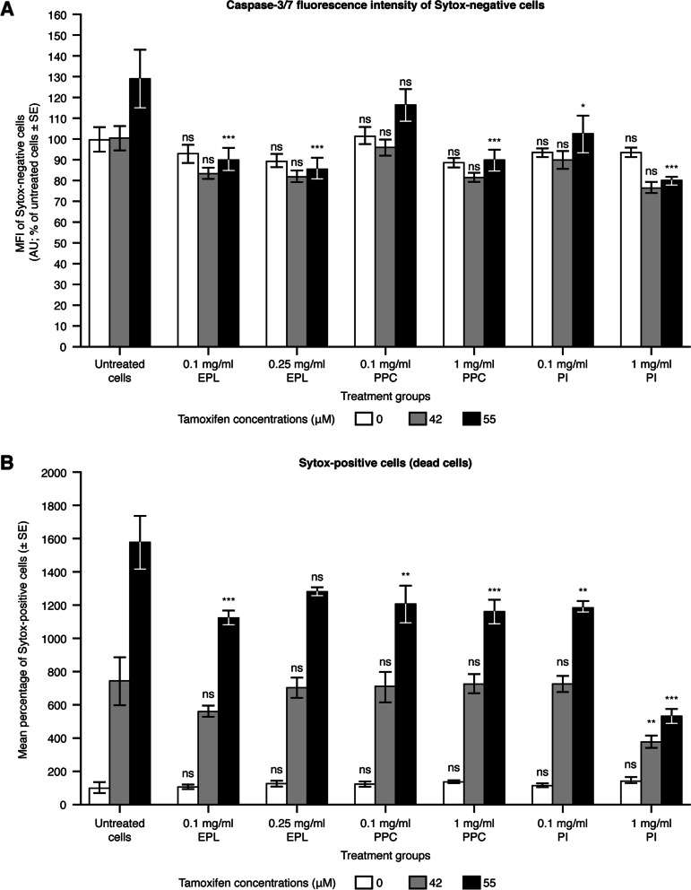
Fig. 2.
Effect of EPL, PPC and PI on apoptosis in the HepG2 cell line. Values shown are mean ± SE (as % of untreated cells) for 2 separate experiments; n = 2 wells for each concentration of each compound per experiment. ns: not significant, *P < 0.05, **P < 0.01, ***P < 0.001 versus untreated cells. Note: for untreated HepG2 cells, 1.43% of cells were found to be Sytox positive (dead cells). Here, these values are presented as percentages, as the results are normalized to untreated cells. AU, arbitrary units; EPL, essential phospholipids; ns, not significant; MFI, median fluorescence intensity; PI, phosphatidylinositol; PPC, polyenylphosphatidylcholine; SE, standard error. Supplementary Table S3 shows the statistical analyses

