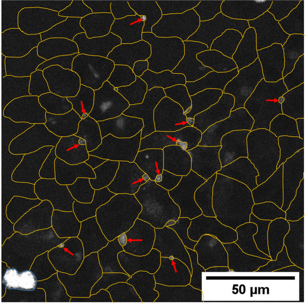
Fig. 5.

Visualization of BSEP in 0.25 mg/ml EPL treated steatotic HepaRG cells. Representative steatotic HepaRG cell cultures treated with 0.25 mg/ml EPL and visualization of BSEP as accumulation of the substrate 7-beta-NBD-taurocholate in canaliculi. Yellow lines show the individual cells. Red arrows highlight some example spots of canaliculi structures with accumulated fluorescent substrate. The blue circled area with high fluorescence intensity is a dead cell where the fluorescence substrate accumulates. BSEP, bile salt export protein; EPL, essential phospholipids
