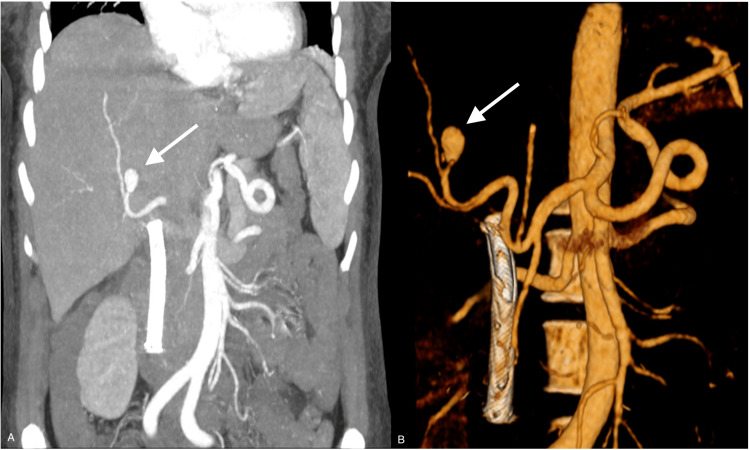
Figure 3. CTA (A) and 3D angiographic study (B) reconstruction of the abdomen showing the presence of a pseudoaneurysm is documented in a segmental branch of the right hepatic artery with a diameter greater than approximately 12 mm (arrows); a biliary stent previously placed in the common bile duct can also be seen.
CTA: computed tomography angiography; 3D: three-dimensional

